Application Note 5: Vertical Translation Option for Density Gradient Sorted Samples
The vertical translation option is available to add to any of our NanoSpectralyzer models (NS1, NS2, or NS3). It allows for the controlled motion of the sample in the vertical direction (45 mm range). In NanoSpectralyzers equipped with the vertical translation option (VTO), the sample cell holder is mounted on a vertical translation stage driven by a computer-controlled stepper motor. This allows automatic scans of fluorescence, absorption, and Raman spectra as a function of position in the sample cell, using the Sequence Acquisition tab of the Main control screen. Such spectral maps are particularly valuable for in situ characterization of layered contents in centrifuge tubes used for density gradient ultracentrifugation sorting of SWCNT samples. The VTO cell holder is designed to accommodate centrifuge tubes with outer dimensions of 13 x 51 mm or 14 x 89 mm (diameter x height), in addition to conventional square 12.5 x 12.5 mm spectrophotometer cells.
|
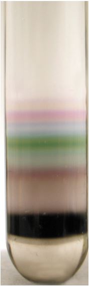
One of the most promising methods for purifying and sorting SWCNTs is density gradient ultracentrifugation (DGU). An aqueous surfactant-suspended SWCNT sample is placed into a centrifuge tube filled with a medium of spatially varying density. Iodixinol is commonly used as the high density water-soluble component in these preparations. After extended high speed centrifugation, it is found that the SWCNTs become spatially stratified and separated according to physical properties. Proper choice of surfactant and density gradient conditions allows even individual (n,m) species to be segregated into distinct layers. Careful fractionation of the centrifuge tube contents then gives structurally sorted SWCNT samples that are extremely useful for a number of applications. A key step in the delicate optimization of DGU parameters (density profile, surfactant choice, centrifugation speed, centrifugation time, etc.) is analyzing the compositional profile of the centrifuge tube after a run. Until recently, this has required painstaking layer-by-layer fractionation of the tube’s contents into many small samples, followed by spectrometric analysis of each fraction to identify its SWCNT composition. Now, however, the NanoSpectralyzer can perform this analysis quickly, conveniently, and noninvasively in the intact centrifuged sample. |
|
Method
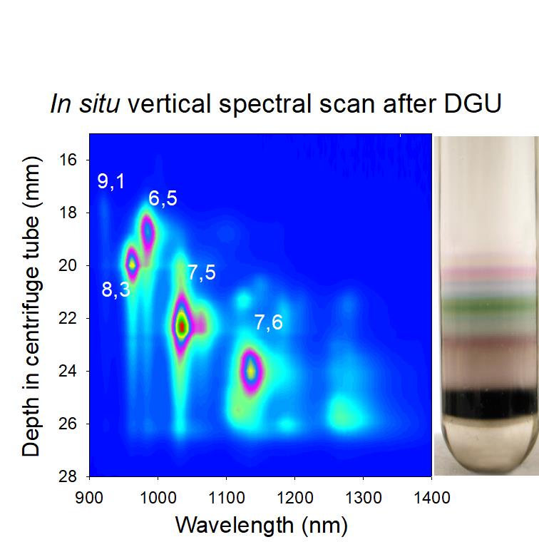
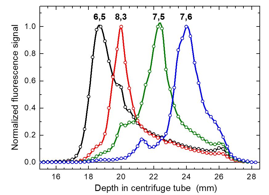
NanoSpectralyzers use coherent and tightly focused laser beams to excite fluorescence from semiconducting SWCNTs. The dimensions probed in the sample are therefore very small. In addition, the NanoSpectralyzer optical design allows fluorescence measurements from cylindrical as well as conventional (rectangular) sample cells. So with a simple adaptation of the sample NanoSpectralyzer sample cell holder, one can use a 14 mm diameter centrifuge tube (compatible with SW41 rotors) as the sample cell. Fluorescence data are acquired in situ, right through the wall of the undisturbed centrifuge tube. Another minor option allows vertical translation of the centrifuge tube relative to the optical axis. Scanning the tube position gives near-IR fluorescence spectra with sub-millimeter vertical resolution. At each vertical position, spectra can be acquired using all three excitation wavelengths and then automatically analyzed to provide a full (n,m) composition map as a function of depth in the tube. This method offers far greater speed and spatial resolution than post-fractionation analysis. The NS1 can also measure absorption spectra as a function of vertical position to monitor non-emissive sample components.
Example data
This contour plot shows emission intensity as a function of wavelength and vertical position in a DGU centrifuge tube. Data collection time was only a few minutes. Although this plot represents emission using only one of the three excitation lasers in the NanoSpectralyzer, it allows simple identification of the major (n,m) species at each vertical position and accurately reveals the extent of physical separation among these species. This plot can also guide fractionation efforts by showing the exact depths from which to collect each species of interest.
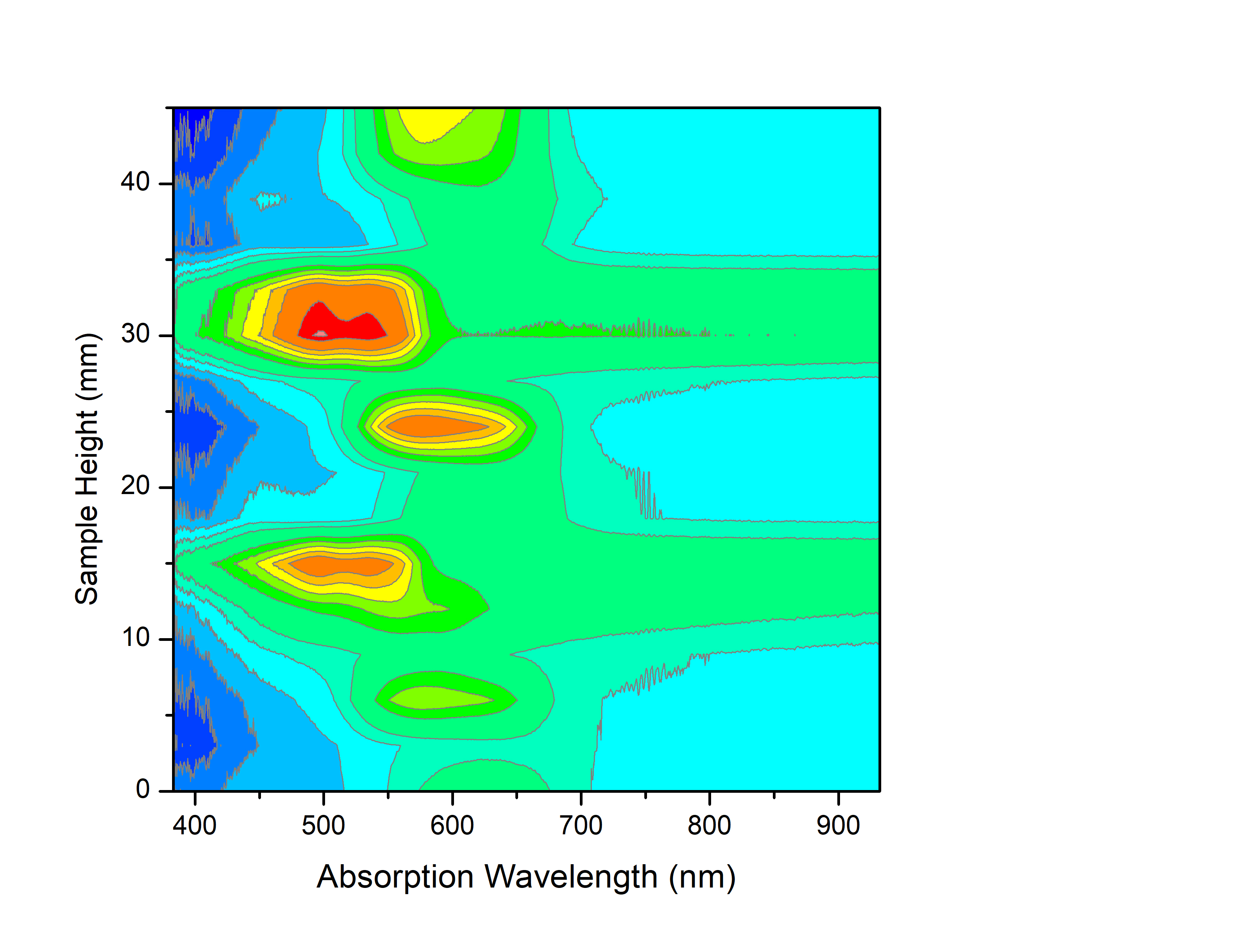
Additionally, the vertical translation option is not restricted to fluorescence data. Below is shown a contour plot of the visible absorption data for a glass tube with stripes drawn on it using different color markers as the glass tube is scanned vertically.